

Your Elgin Eye Doctor


Our Eye Care Services

Comprehensive Eye Exams
From updating your eyeglasses prescription to detection and treatment of eye diseases, comprehensive eye exams are important for continued visual health.

Cataract Surgery Co-Management
If cataracts go untreated, they can cause total blindness in the affected eye. We can help with co-management of your cataract removal surgery.

Dry Eyes
Do you have dry, itchy, gritty-feeling eyes? Dry eye syndrome is a very common condition. We offer dry eye treatments at our eye care clinic.
Children with myopia are at higher risk for potentially sight-threatening vision problems. Save your child’s vision with myopia management.
LASIK and refractive surgery are great ways to say goodbye to contacts and glasses for good. Find out if you’re a good candidate.
Over 50% of people living with glaucoma don’t know they have the disease as it shows no symptoms. Early detection and treatment can prevent blindness.
During a contact lens exam, your eye doctor will check if you are a good candidate for contacts and find the best type of contacts for your needs..
Eye emergencies such as severe pain in the eye, or foreign objects puncturing the eye require immediate attention. Contact us immediately.
Astigmatism can cause vision problems such as blurred or double vision at all distances. Find out what you can do to correct astigmatism and see your best.
Macular degeneration can cause severe central vision loss if not detected and treated early on. We can help preserve your vision.
The ups and downs of diabetes can be difficult to navigate. How can diabetes affect your eyes? How can you and your eye doctor keep them healthy?
Presbyopia is a normal part of aging. If you’re past 40 and notice you can’t read up close, our eye doctors may be able to help.
Scleral lenses are a great option for those with severe dry eye or abnormally-shaped corneas. Is this comfortable alternative to traditional contacts right for you?
Orthokeratology is a non-invasive vision correction alternative to LASIK. Put in the specialty lenses while you sleep, and experience better vision in the morning.
Eyeglasses & Frames

Designer Frames
Our extensive optical section offers a wide variety of eyeglass frames in every style, material & design. Come visit us today to see for yourself!
Our expert optical team can find just the right pair of glasses for you to be confident and look your best.
Lens coatings improve visual comfort, make it easier to clean your glasses and ensure your lenses last longer. Coatings include anti-scratch, anti-reflective, photochromatic and UV / blue light filters.
Contact Lenses

Contact Lens Fitting
We offer a wide range of contact lens options from dailies, monthly to multifocal contact lenses for crystal clear vision and superior comfort.
During a contact lens exam, your eye doctor will check if you are a good candidate for contacts and find the best type of contacts for your needs.
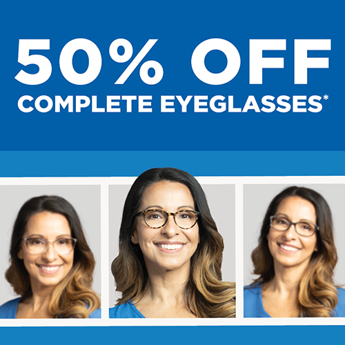
Buy One, Get One 50% Off Eyeglasses.
*Requires purchase of complete prescription pairs, including frame and lenses. Discount applied to complete pair of equal or lesser value. Does not include sunglass frames,Barton Perreira, Cartier, Cazal, Chanel, Cutler and Gross, Dior, Dita Lancier, Fendi, Gucci, ic!Berlin, l.a. Eyeworks, Maui Jim, Mykita, Nifties, Oakley, Oliver Peoples, Persol, Ray-Ban, Robert Marc, Salt, Salvatore Ferragamo, Skaga, Silhouette, Tom Ford, WOOW, accessories, contact lenses, or medical procedures. Cannot be combined with any other discounts, promotions, or insurance plans. Not valid on previous orders. Other restrictions may apply. See practice for full details. Offer valid 04/08/2024-06/16/2024. 24AEG-729313


Location & Opening Hours
We are at the corner of Weld and Randall south of Rt 20. We are close to Marcus movie theater and Old Republic Kitchen.
- Monday 9:00 am - 6:00 pm
- Tuesday 9:00 am - 7:00 pm
- Wednesday 9:00 am - 6:00 pm
- Thursday 9:00 am - 7:00 pm
- Friday 9:00 am - 6:00 pm
- Saturday 8:00 am - 2:00 pm
- Sunday Closed